Figure 3.
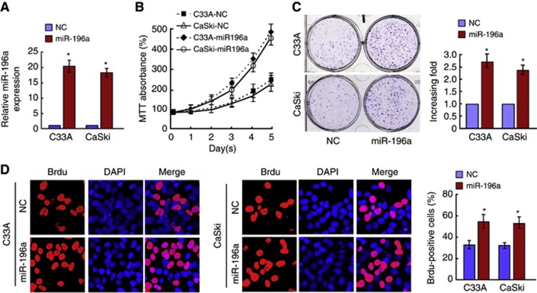
miR-196a upregulation promotes proliferation of cervical cancer cells. (A) Expression of miR-196a in C33A and CaSki cells transfected with miR-196a mimics (miR-196a) by real-time PCR. (B) Effects of miR-196a overexpression on the growth of cervical cancer cells C33A and CaSKi. 3-(4,5-Dimethyl-2-thiazolyl)-2,5-diphenyl-2H-tetrazoliumbromide assays showed that miR-196a-transfected C33A and CaSKi cells proliferated more rapidly than the control cells. (C) Representative micrographs (left) and quantification (right) of crystal violet-stained cell colonies. (D) Representative micrographs (left) and quantification (right) of BrdU–incorporating cells after transfection with miR-196a or negative control (NC). Error bars represent mean±s.d. from three independent experiments. *P<0.05.

