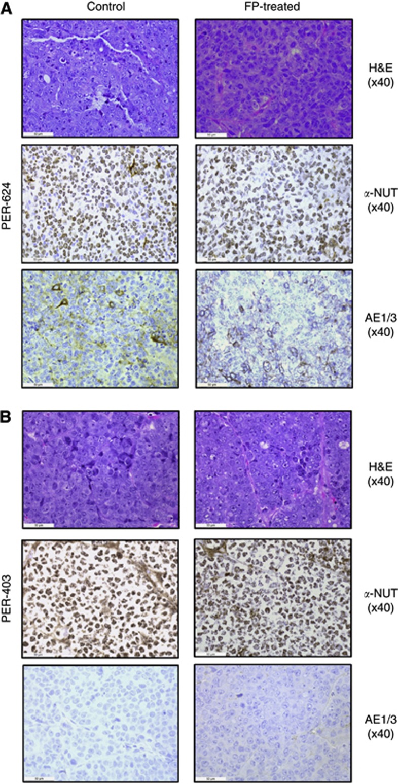
Figure 4.
Comparison of control and FP-treated tumours from (A) PER-624 or (B) PER-403 xenografts. Images show haematoxylin & eosin (H&E) staining, IHC for NUT (α-NUT), and cytokeratin (AE1/3), with brown immunopositive stain and blue haemotoxylin nuclear counterstain. In each case, tumours were poorly differentiated, with cells demonstrating large nuclei and prominent nucleoli, scant cytoplasm with little cytokeratin and extensive speckled nuclear staining for NUT. There was no discernable difference between tumours from control and FP-treated xenografts in either line.

