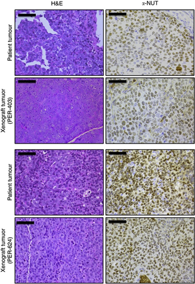
Figure 5.
Histological comparison of NMC xenograft tumours (PER-403 and PER-624) with the primary patient tumours from which each cell line was derived; left panel, haematoxylin & eosin (H&E); right panel, anti-NUT IHC (α-NUT, brown; haematoxylin counterstain, blue), magnification × 40, bars 50 μm. All tumours were poorly differentiated, with cells demonstrating large nuclei with prominent nucleoli, scant cytoplasm and extensive speckled nuclear staining for NUT. A small but similar proportion (∼10%) of cells in each tissue was negative for NUT. Alveolar structures are visible in the primary tumour (top panel) used to derive PER-403.

