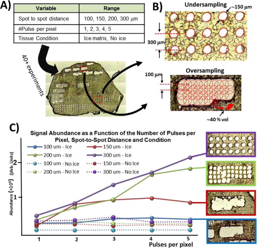
Figure 3.
A) Combination of parameters tested on a liver tissue section (50 µm). For each condition, abundance of selected peaks were computed and optical image of the tissue section captured. B) Undersampling vs oversampling techniques and how overlapping pixels can lead to better spatial resolution. C) Optical images of ablated tissue and corresponding average abundance for different spot-to-spot distances and number of pulses per pixel. As expected, multi-shot imaging of tissue sections without an ice matrix did not result in greater signal since all material is ablated after first pulse (also confirmed with optical images). Greater signal can be achieved using ice as a matrix where it becomes a trade-off between spatial resolution and sensitivity.

