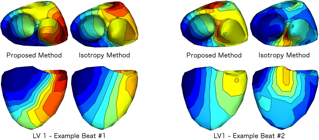
Fig. 5.
Comparison of the Proposed Method to the Isotropy Method: We applied our proposed method and the isotropy method to all beats from all pacing locations, using the same transmural gradient regularization in both methods. As an example, we show two views (top: endocardial, bottom: epicardial) of isochrone maps of results from each method applied to two consecutive beats paced from the same location (subject 1, LV 1). As in Fig. 3, maps were normalized to have the same duration, thus the earliest activation time is blue and the latest activation time is red in each map (see Fig. 3 for colormap). Example beat #1 is a case where results from the isotropy method and our proposed method were similar. Example beat #2 shows the next subsequent beat from the same pacing site, where the results from the two methods were dissimilar.

