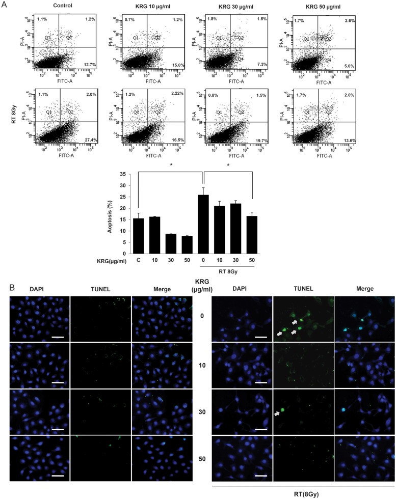
Figure 5:
Effect of KRG on radiation-induced apoptosis in HaCaT cells. (A) In order to analyze quantitatively the effects of KRG on radiation-induced apoptosis, we used the Annexin V-FITC and PI assay. The percentage of apoptotic cells of those treated with radiation in the absence or presence of KRG were indicated above (upper). The percentage of apoptosis in each fraction was expressed as a graph (lower). KRG significantly decreased apoptosis at a dose of 50 µg/ml. The data represent the mean ± SD of four independent experiments. *P < 0.05. (B) Apoptosis in HaCaT cells was identified by the TUNEL method using an in situ cell detection kit. After cell monolayers reached 60–70% confluence, the cells were exposed to a medium with radiation (8 Gy) in the presence of 0, 10, 30 or 50 µg/ml of KRG. The cells were then incubated with 50 µl of TUNEL reaction mixture (TdT and fluorescein-dUTP) and stained with Hoechst 33258 (5 µg/ml). The stained cells (arrow) were observed by fluorescence microscopy. The TUNEL assay confirmed that radiation induced TUNEL-positive cells (arrow) and KRG decreased TUNEL-positive cells. Scale bar = 50 µm.

