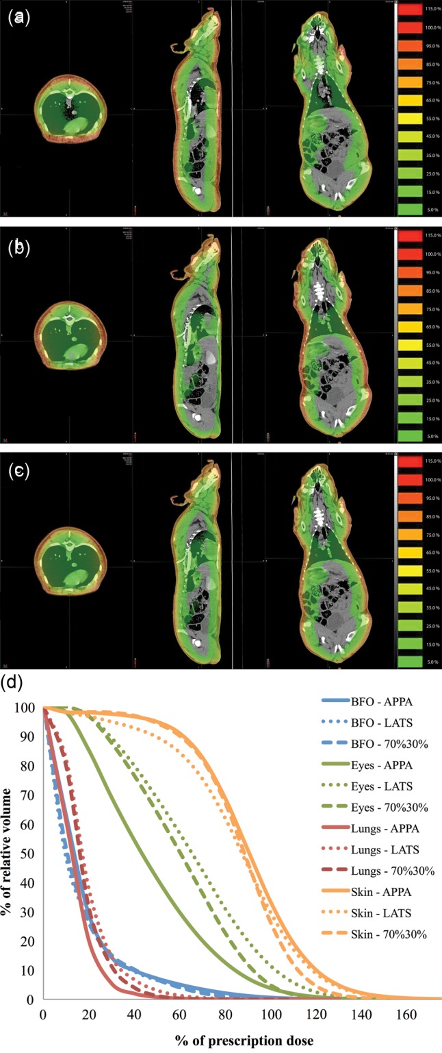
Fig. 5.

Dose distributions overlaid on Yucatan minipig CT images for 6 + 12 MeV electron irradiations for (a) opposed anterior–posterior beams (APPA), (b) evenly weighted lateral opposed (LATS), and (c) a combination of 70% LATS with 30% APPA beams. The images shown are the transverse, sagittal and coronal planes. (d) DVHs of eyes, lungs, BFO and skin for the three beam orientations.
