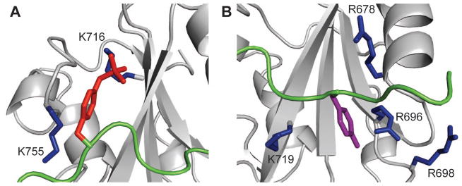
Figure 5.

Ribbon representation in close-up view of the two binding pockets of Vav1 SH2 (grey) ligated with SykLB-YpY (green). A: The specificity pocket occupied by phosphorylated pY346 (red sticks). K716 and K755 are highlighted with sticks. The key interaction for recognition of phosphotyrosine in the specificity pocket is with K716. B: The pTyr pocket occupied by unphosphorylated Y342 (purple sticks). Important residues are shown with blue sticks.
