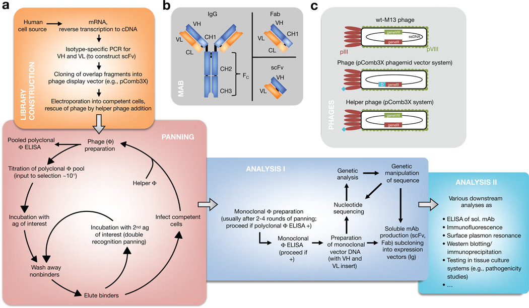
Figure 1. Antibody phage display (APD).
(a) Diagram showing how APD is performed. (b) Interrelatedness of Ig (here, IgG), single-chain variable fragment (scFv), and Fab. Importantly, both scFv and Fab reflect the original specificity of the Ig they are derived from because variable heavy (VH) and variable light (VL) chains form the interface with the antigen. (c) Phage display systems are derived from wild-type bacteriophage of Escherichia coli. Monovalency of the displayed protein (turquoise) ensures selection for high-affinity mAbs in phagemid vector-based systems (e.g., pComb3X). Helper phages display a fusion protein as well after propagation from phage-infected cells and are also selected during panning (but they do not possess the sequence of the displayed protein within). A defective helper phage origin of replication ensures preferential production and packaging of the desired phage particles, requiring addition of new helper phage in every panning round.

