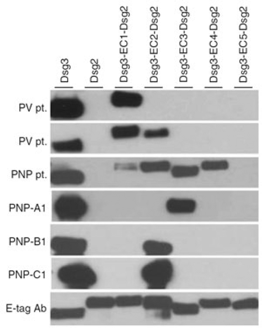
Figure 4. Three anti-Dsg3 scFvs (PNP-A1/B1/C1) isolated from a PNP patient bind to the EC2 and EC3 domains of Dsg3 in immunoprecipitation/immunoblot analysis of the patient’s and a PV patient’s sera.
The PNP patient’s sera recognized Dsg3 epitopes EC1–4, but the mAbs derived by phage display recognized only EC2 and EC3. In classic PV patients, the main epitopes are EC1 and EC2. These findings also demonstrate that antibody phage display may miss some pathologic clones (here, such mAbs that target EC1 and EC4) for various reasons, as discussed in the last paragraph of this article. Dsg, desmoglein; PNP, paraneoplastic pemphigus; PV, pemphigus vulgaris; scFv, single-chain variable fragment. Reprinted with permission from Saleh et al. (2012).

