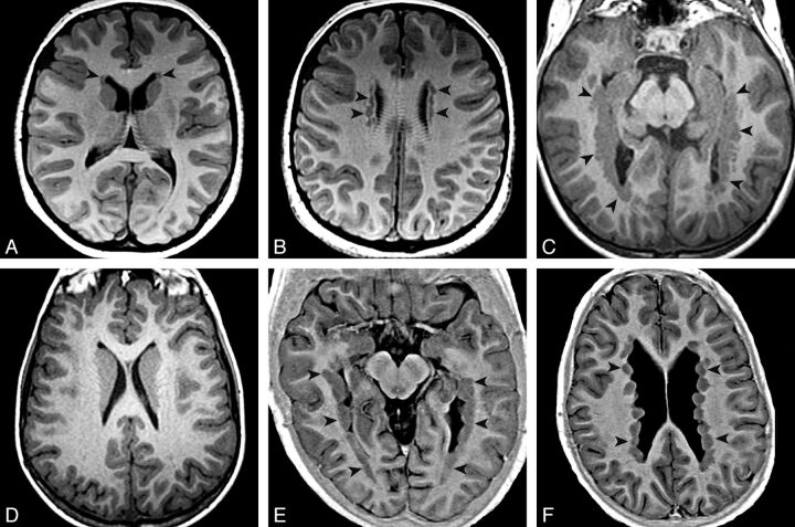
Fig. 1.
PNH main patterns evaluated with T1 inversion recovery–weighted image. Arrowheads show locations of heterotopia. Axial images show an aPNH pattern with nodules restricted to the frontal horns (A) and bodies (B). Axial images demonstrate a pPNH pattern. Nodules are lining trigones and temporal and occipital horns bilaterally (C), sparing the frontal horns and bodies (D). E and F, Axial images show a dPNH pattern with nodules lining all of the walls of the lateral ventricles.

