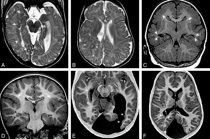
Fig. 2.
pPNH and malformations of cortical development. Axial T2 FSE images show PNH lining the wall of the right temporal and occipital horns and trigone (arrowheads in A) and a large region of subcortical heterotopia in the right frontoparietal region (asterisks in B). C, Coronal T1 inversion recovery–weighted image demonstrates bilateral PNH in the temporal horns (arrowheads) and bilateral parietal pachygyria (arrows). D, Coronal inversion recovery–weighted image shows right temporal PNH (arrowheads) and bilateral perisylvian polymicrogyria (white arrows). E and F, Axial T1 inversion recovery–weighted images demonstrate bilateral pPNH (arrowheads) and a left occipital schizencephaly (asterisk in E). White arrows in F show the thickness of the WM band measured between the most posteromedial margin of Sylvian fissure and the lateral border of the ventricular trigone. In this case, it was diminished (5 mm).

