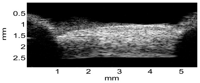
Figure 5.

A representative ultrasound image of the porcine posterior sclera noted with the actual physical dimensions, showing the typical intrinsic ultrasound speckle patterns, adequate sample flattening, and the locations of sample mount (shadows on both sides).
