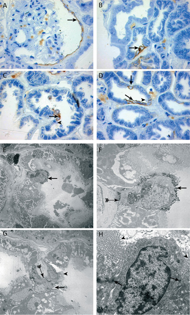
Figure 3.
Ultrastructural analysis of CD24 immunolabelled cells in semi-thin and ultrathin sections. (A – D) CD24 (brown staining) is expressed by the parietal epithelial cells of Bowman’s capsule (A, arrow) and a few proximal tubular cells (B – D, arrows). An abrupt termination of the brush border was visible on cells adjacent to the CD24-positive cells (C, D, arrowheads). Immunoelectron (E – G) and transmission electron microscopy (H). The morphology of CD24-positive cells (black outlining, E – G, arrows) differed from that of normal proximal tubular cells. CD24-positive cells contained less cytoplasm, fewer mitochondria, and the brush border was mostly rudimentary or absent compared with the brush border of adjacent CD24-negative cells (G, H, arrowheads). Of note, some CD24-positive cells contained a brush border. Panel G shows two CD24-positive cells adjacent to each other, one of which lacks a brush border (G, arrow) and the other does not (G, arrowhead). In addition, the basolateral membrane lacked the typical invaginations (F, G, arrow with tail). Electron microscopy revealed desmosomes between the two morphologically different proximal tubular cells (H, arrowheads).

