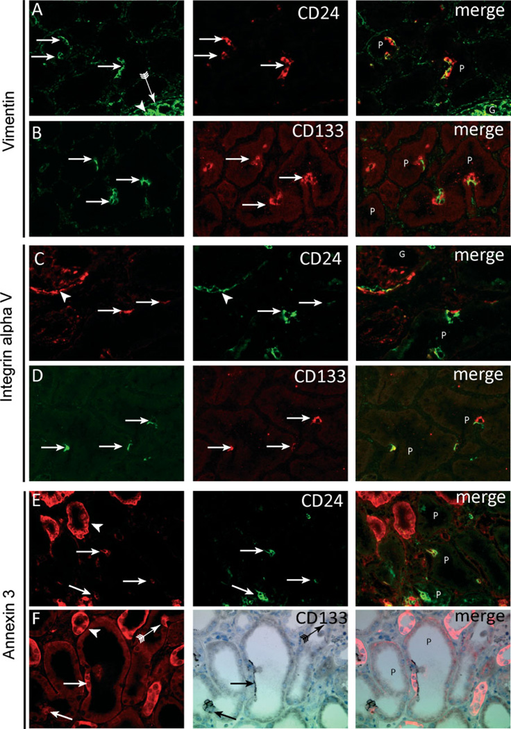
Figure 4.
CD24- and CD133-positive cells exhibit distinct marker expression. In contrast to neighbouring cells, CD24- and CD133-positive proximal tubular cells (A – F, arrows) show expression of vimentin (A, B), integrin αV (C, D), and annexin 3 (E, F). Vimentin (A, B) was strongly expressed by podocytes (A, arrowhead) and parietal epithelial cells (A, arrow with tail) but also showed co-localization in the CD24- and CD133-positive proximal tubular cells (A, B, arrows, respectively). Integrin αV (C, D) was expressed by the parietal cells (C, arrowhead) and the CD24- and CD133-positive proximal tubular cells (C, D, arrows, respectively). Annexin 3 (E, F) was expressed in distal tubuli (arrowhead) and in the CD24- and CD133-positive proximal tubular cells (arrows). The arrow with tail in panel F shows an annexin 3 cell negative for CD133. G = glomerulus; P = proximal tubules.

