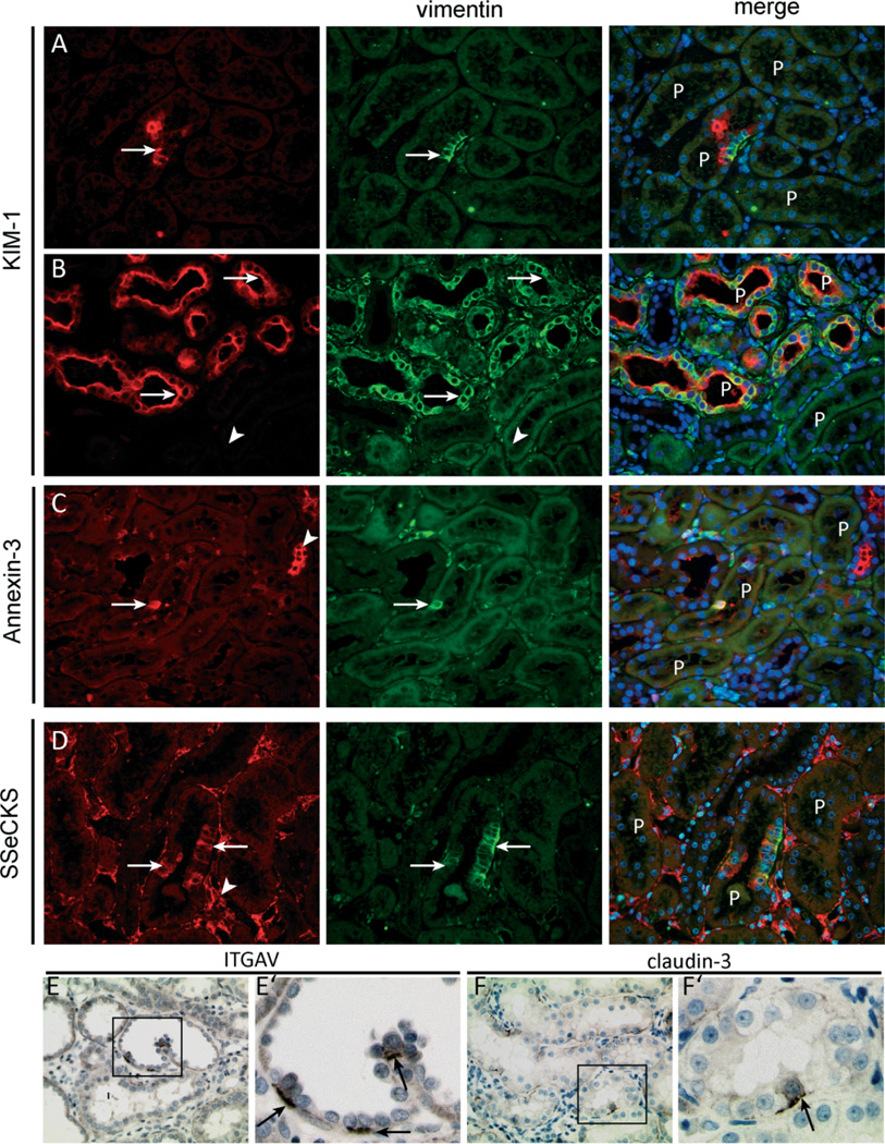
Figure 8.
Vimentin-positive proximal tubular cells exhibit distinct marker expression. (A, B) The vimentin-positive cells in the injured rat kidneys co-expressed KIM-1. Vimentin and Kim-1 co-expression was seen in single cells or small groups of cells (A, arrow) but also in complete proximal tubular structures in segments of the kidney with more advanced tubular injury (B, arrows). Adjacent normal-appearing tubuli were mostly negative for both markers (B, arrowhead). (C) Within proximal tubuli, vimentin co-localized with annexin 3 (arrow). Annexin 3 was also expressed in other tubular structures (arrowhead). (D) Vimentin-positive proximal tubular cells co-expressed SSeCKS (arrows), which was also expressed by endothelial cells (arrowhead) and parietal cells (not shown). (E, F) Within the affected rat kidneys, both integrin αV and claudin-3 were expressed by a few proximal tubuli in a scattered pattern. E’ and F’ show a zoomed picture of the insets in E and F, respectively, and show mostly basolateral staining of both markers in a subset of the proximal tubular cells.

