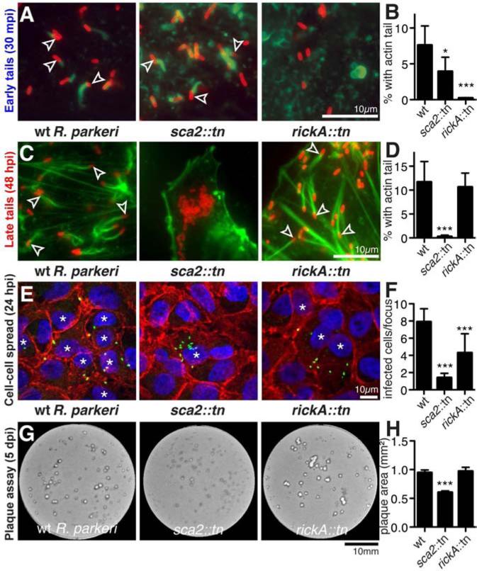
Figure 3. Early actin-based motility requires RickA and late motility requires Sca2.
(A, C) HMEC-1 cells infected with wild type or mutant R. parkeri for (A) 30 min or (C) 48 h, stained with Alexa Fluor 488-phalloidin to label actin and anti-Rickettsia antibody. Arrowheads indicate bacteria with an actin tail. (B, D) Percentage of wild type or mutant R. parkeri with actin tails in HMEC-1 cells infected for (B) 30 min or (D) 48 h. Data are mean ± SD of 5 random fields of view in each of three independent experiments performed in duplicate. (E) Infectious foci formed by wild type or mutant R. parkeri in A549 cells, stained with DAPI (blue), anti-β-catenin antibody (red) and anti-Rickettsia antibody (green) (* infected cells). (F) Number of infected cells per focus for strains in (E). Data are mean ± SD for 10 foci. (G) Plaques formed by wild type or mutant R. parkeri in Vero cells 5 dpi. (H) Plaque area for strains in (G). Data are mean ± SEM of data from three independent experiments. *p<0.05, *** p<0.001 indicate significant variation of mean from wild-type values by ANOVA with Bonferroni's multiple comparison test. See also Figure S3.

