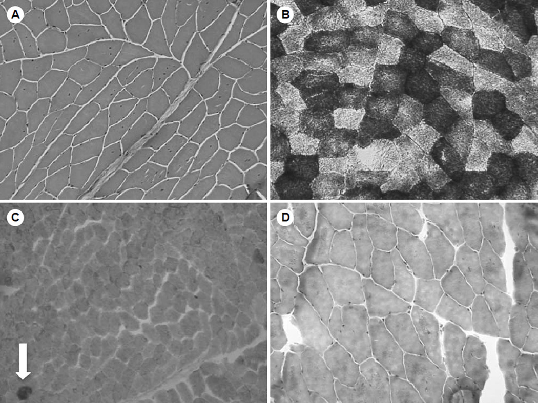
Fig. 2.
Immunohistochemical analysis of biceps muscle. A) Hematoxylin and eosin staining shows many muscle fibers with central nuclei. B) Staining with nicotinamide adenine dinucleotide hydride demonstrates that many muscle fibers have “motheaten” appearance. C) Staining for cytochrome C oxidase and succinyl dehydrogenase demonstrates 2 cytochrome C oxidase–negative ragged blue fibers (arrow) on low power. D) Modified Gomori trichrome stain demonstrates no obvious ragged red fibers.

