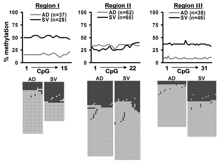
Figure 4.
Comparison of CpG methylation patterns within the Mest promoter in DNA isolated from the adipocyte (AD) and stromal-vascular (SV) fractions of adipose tissue show enhancement of hypomethylated alleles in genomic regions I and III of adipocytes. Pooled inguinal fat from B6 mice fed a chow diet were sub-fractionated into fractions that were enriched for mature adipocytes and SV cells. The graphs show the temporal frequency of CpG methylation (% methylation) of genomic regions I, II and III for each cellular fraction. A minimum of 28 clones were sequenced and analyzed for each of the cellular fractions and the individual alleles showing unmethylated (white boxes) and methylated (gray boxes) CpGs within each genomic region are shown under the line graphs.

