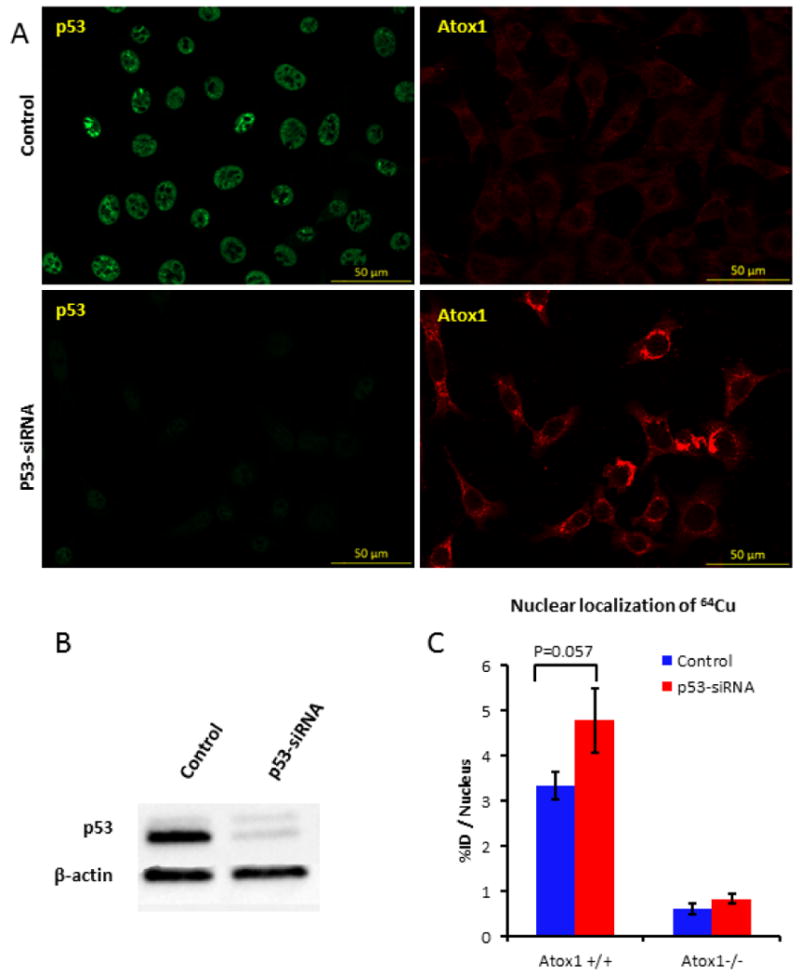
Figure 4.

Effect of p53 knock-down on Atox1 expression in MEF Atox1+/+ cells. (a) Confocal images of MEF Atox1+/+ cells after KD of p53 using p53-siRNA. Cells were seeded on glass coverslips, treated with 20 pmol p53-siRNA for 48 h and fixed with 4% PFA. p53 (green) was detected with Mouse Anti-Human p53 antibody and Atox1 (red) was detected with Goat Anti-mouse HAH1 antibody. (b) Western blot showing p53 knock-down in MEF Atox1+/+ cells. (c) Nuclear localization of [64Cu]copper acetate in MEF Atox1+/+ and Atox1-/- cell lines before and after p53 knock-down (n=3). These results are representative of two independent experiments.
