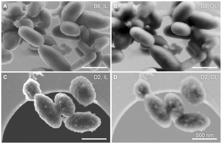
Figure 4. SEM images of freeze-dried C57BL/6J (B6) and DBA/2J (D2) melanosomes.
The left column contains images recorded in the in-lens detector configuration (IL) and the right column shows micrographs acquired in the out-lens configuration (OL). A direct comparison shows that B6 melanosomes (A and B) have a rather smooth and featureless surface, whereas the D2 organelles (C and D) have an irregular surface. The samples were subjected to the identical preparation protocol. All scale bars represent 500 nm.

