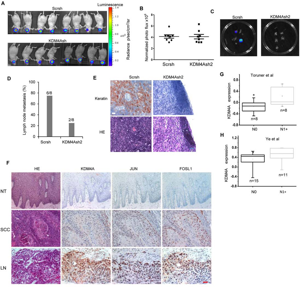
Fig 7. KDM4A is required for human SCC lymph node metastasis in vivo.
(A and B) Xenograft tumors derived from SCC/MET/Scrsh cells (n = 8) or SCC23/MET/KDM4Ash2 cells (n = 8) in the oral cavity of mice was assessed by bioluminescent imaging for 14 or 20 days, respectively. Data are representative of two independent experiments. (C) Lymph node metastasis resulting from tumors in (A) assessed ex vivo by bioluminescent imaging. (D) Number and percentage of mice with lymph node metastasis from xenograft tumors described in (A). **P< 0.01, Fisher’s exact test. (E) Histochemical detection of SCC lymph node metastases from (D) using an antibody against Pan-keratin. H&E staining provided for histology. Images are representative of −6 metastatic samples derived from SCC23/MET/Scrsh cells and 6 non-metastatic samples from SCC23/MET/KDM4Ash2 cells. Scale bar = 50 µm. (F) Histochemical detect ion of KDM4A, JUN, and FOSL1 abundance in human SCC tissue (SCC) and SCC lymph node metastases (LN) compared with adjacent normal epithelial tissue (NT). Images are representative of 35 (NT), 68 (SCC), and 34 (LN) samples. (G and H) Oncomine analysis of KDM4A mRNA abudance in human primary SCC with lymph node metastasis (N1+) compared with human primary SCC without metastasis (N0) reported in (G) Toruner et al. (32) and (H) Ye et al. (33). *P<0.05, unpaired two-tailed Student’s t-tests.

