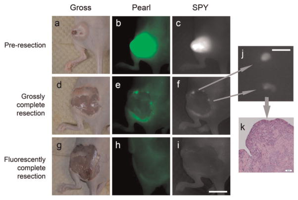
Figure 5.
Detection of microscopic residual disease. Uptake of near-infrared panitumumab-IRDye800 before resection (a–c), after grossly complete resection (d–f), and after fluorescently complete resection (g–i). Fluorescence imaging of microscopic residual disease with the SPY (j) is confirmed by histology with hematoxylin and eosin staining (k). Scale in (i) is 10 mm. Scale in (j) is 3 mm.

