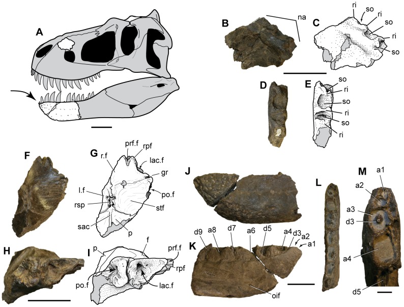
Figure 3. Nanuqsaurus hoglundi, holotype, DMNH 21461. A.
. Reconstruction of a generalized tyrannosaurine skull, with preserved elements of holotype shown in white. Arrow points to autapomorphic, reduced, first two dentary teeth. B–E. Photographs and interpretive line drawings of right maxilla piece in medial (B, C); and dorsal (D, E) views. F–I. Photographs and interpretive line drawings of partial skull roof in dorsal (F, G); and rostrolateral (H, I) views. J–M, partial left dentary in lateral (J); medial (K); dorsal (L) views; and close-up of mesial alveoli in dorsal (M) views. Abbreviations: a, alveolus, with number indicating position in tooth row; d, dentary tooth, with number indicating position in tooth row; f, frontal; gr, orbital groove; lac.f, lacrimal facet of frontal; l.f, left frontal; na, nasal contact surface; oif, oral intramandibular foramen; p, parietal; po.f, postorbital facet of frontal; prf.f, prefrontal facet of frontal;. r.f, right frontal; ri, ridge separating sockets in nasal articulation of maxilla; rpf, rostral process of frontal between prefrontal and lacrimal facets; rsp, rostral spur of parietal; sac, sagittal crest; so, socket for nasal articulation of maxilla; stf; supratemporal fossa. Gray fill indicates missing bone or broken bone surfaces and cracks. Scale bar in A equals 10 cm. Scale bars in B–L equal 5 cm. Scale bar in M equals 1 cm.

