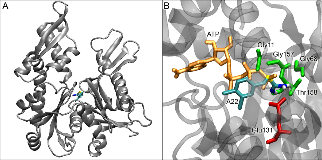
Figure 2. A22 docking in the MreB nucleotide cleft.
(A) Docking site of A22 in the nucleotide-free form of MreB. (B) Details of the binding site of A22 in MreB with residues within 2.5 Å of A22 shown with their sidechains. ATP is shown in its crystal position, illustrating the clashes that would occur between its beta and gamma phosphates and A22.

