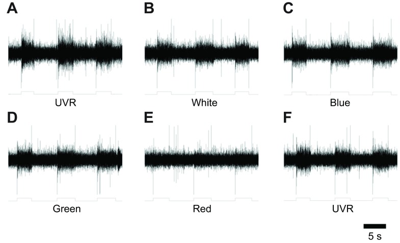
Fig. 4.
Groups of photoreceptors responded to UVR and a range of visible wavelengths. A suction electrode was placed upon the cut end of the DB cephalic nerve that had been severed from the supra-esophageal ganglion. Light from the LED wands was directed at the head and held by hand for a few seconds. Massed photoreceptor responses were complex and slowly adapting as expected (Kretz et al., 1976). The output from a phototransistor (shown below each trace) indicates the timing of the light stimulus and all were delivered at saturating intensities. In each record there was also an instantaneous transient associated with turning the light on (downward) and off (upward). (A) Response to UVR. (B) Response to white light. (C) Response to blue light. (D) Response to green light. (E) Response to red light. (F) Second exposure response to UVR.

