
Figure 1. The neurovascular bundle (NVB) provides a niche for quiescent stem cells.
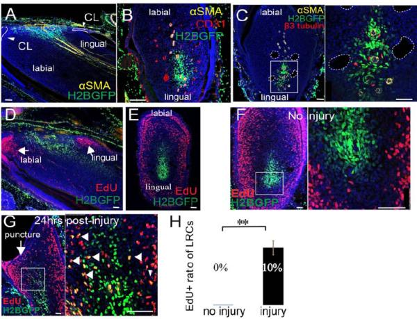
A-B. Sagittal (A) and cross sections (B) of WTH mouse incisors chased for one month (LRCs appear green due to H2BGFP), after αSMA (yellow) and CD31 (red) immunohistochemical staining. In the sagittal sections, the apical region of the incisor is oriented to the left side. In the cross sections, the labial side of the incisor is oriented to the top. αSMA labels arteries. CD31 labels all vasculature. CL, cervical loop. C. β3-tubulin (red) and αSMA (yellow) staining of cross sections of chased WTH mouse incisors. β3-tubulin labels nerves. Boxed area is shown magnified to the right. Dotted white lines outline veins. D-E. Sagittal (D) and cross (E) sections of chased adult WTH mouse incisors treated with EdU (red). F-G. Chased WTH mouse incisors treated with EdU (red) without (F) or with (G) injury to the tooth. Arrow indicates injury site (puncture). Arrowheads indicate double labeling (yellow) of LRCs and EdU incorporation. Boxed areas are shown magnified to the right. Nuclear DAPI staining is in blue. H. Quantification of LRCs incorporating EdU before and after injury, as shown in F and G. Values are plotted as mean ±SEM (n=5, at least 500 cells were counted in each sample; **, p<0.01. n=4). Scale bars, 100μm.