
Figure 2. Gli1+ cells surrounding the NVB support tissue homeostasis and injury repair.
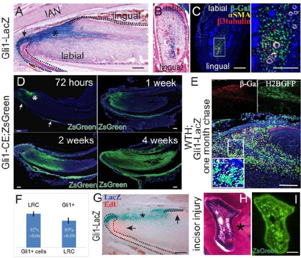
A-C. LacZ staining (blue) of sagittal (A) and cross (B) sections; β-gal (green), αSMA (yellow), β3-tubulin (red) and DAPI (blue) immunohistochemical staining of adult Gli1-lacZ incisor (C). αSMA and β3-tubulin label arteries and nerves, respectively. Boxed area in C is shown magnified to the right. Asterisk indicates Gli1 activity in the mesenchyme. Arrow indicates Gli1 activity in the epithelium. Dotted lines outline cervical loop dental epithelium. D. Time course of Gli1+ cell lineage tracing (green) in adult Gli1-CE;ZsGreenflox mice after tamoxifen induction. Asterisk indicates incisor mesenchyme derived from Gli1+ cells. Arrows indicate Gli+ cell derivatives in the epithelium. E. β-gal staining in chased adult WTH;Gli1-LacZ tetra-transgenic mouse incisors shows Gli1+ and LRC colocalization. Red β-gal staining indicates Gli1 expression. LRCs appear green from H2BGFP. The boxed area is enlarged in the inset. Yellow cells are double stained for Gli1+/LRCs. F. Quantification of results from panel E. 95%±0.06 of Gli1+ cells are LRCs, whereas 80%±0.09 of LRCs are Gli1+. Values are plotted as mean ±SEM (n=4). G. LacZ (blue) and EdU (pink) staining of incisors from 6-week-old Gli1-LacZ mice. EdU was injected two hours before collecting samples. Dotted lines outline cervical loop dental epithelium. H-I. H&E staining (H) and fluorescent image (I) of incisor cross sections from 4- to 6-week-old Gli1-CE;ZsGreen mice after incisor injury. Asterisk indicates reparative dentin formation. Images from adjacent sections show the contribution of Gli1+ cells to reparative dentin formation. Scale bars, 100 μm