
Figure 3. Sensory nerves provide Shh to Gli1+ cells. Denervation disrupts the incisor MSC niche and causes abnormal phenotypes.
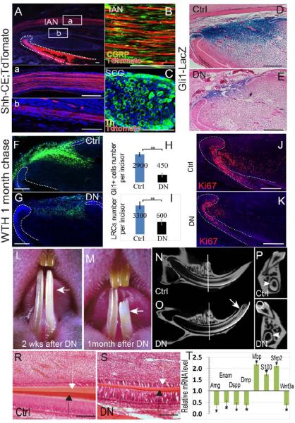
A. Adult Shh-CE;TdTomato mouse incisors 2 weeks after tamoxifen induction. Boxed areas (a) and (b) are shown magnified below. Note the Shh expression (red) in the inferior alveolar nerve (IAN) (a) and nerve bundles accompanying the artery (b). Dotted lines outline the dental epithelium. DAPI staining is in blue. B. Sensory nerve marker CGRP staining (green) of the IAN in adult Shh-CE;TdTomato mouse incisors. C. Th staining (green) of the superior cervical ganglion (SCG) in adult Shh-CE;TdTomato mouse incisors. The SCG is negative for Shh activity (red). Th staining labels sympathetic neurons. D- E. LacZ staining (blue) of control (Ctrl) or denervated (DN) Gli1-lacZ incisors indicates significantly reduced Gli1 activity after denervation. Dotted lines indicate the cervical loop epithelium. F-G. LRC (green) of control (Ctrl) or denervated (DN) WTH incisors one month after chasing. Dotted lines outline the dental epithelium. H-I. Quantification of results from panels D-E (H) and F-G (I). Values are plotted as mean ±SEM (**, p<0.01; n=5). J-K. Ki67 staining shows fewer proliferating cells in the mesenchyme of denervated incisors (K) as compared with control (sham-operated) incisors (J). L-M. Denervated incisors (arrow) turn chalky white 2 weeks after surgery (L; n=20) and fracture within a month (M; n=14). N-Q. Longitudinal micro-CT images of sham-operated (control) incisor (N) and denervated incisor (O). Arrow indicates the fracture site. Cross sections (P, Q) were sampled at comparable positions, indicated by white lines in N-O. Arrowheads indicate the dentin wall of the incisors. R-S. HE staining of control (Ctrl) and denervated (DN) incisor longitudinal sections after one month. Note the reduced thickness of the enamel (black arrow) and dentin (white arrow). T. Real-time PCR data of indicated genes in denervated incisors compared to control incisors. Values are plotted as mean ±SEM (*, p<0.05; n=4). Scale bars, 100 μm.