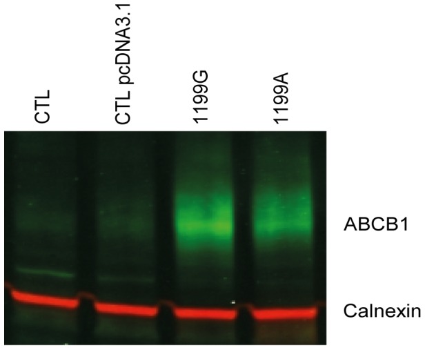
Figure 2. ABCB1 expression analysis by western blot.

(A) Untransfected (CTL) HEK293, (B) transfected with empty plasmid (CTL pcDNA3.1) HEK pcDNA3.1 (C) HEK1199G and (D) HEK1199A cells lyzed. Proteins extracts were analyzed by western blot using two color odyssey system with anti-ABCB1 (green) and anti-calnexin (red) antibodies as described in materials and methods.
