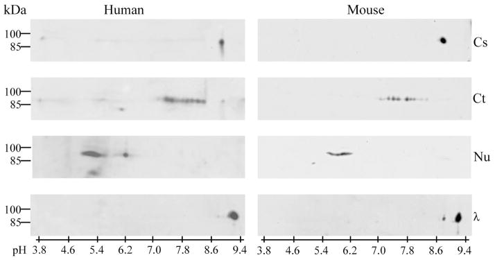
Fig. 4.

Brain cytosolic (Cs), cytoskeletal (Ct), and nuclear (Nu) fractions of human (left panel) and mouse (right panel) were denatured prior to separation by 2-D electrophoresis, and DYRK1A was detected in Western blots with mAb 8D9. In cytosolic fraction, DYRK1A was observed exclusively at pI 8.7, and after dephosphorylation with λPPase (λ), its pI shifted to 9.0. In the cytoskeletal and nuclear fractions of both human and mouse brains, different patterns were observed: several discrete focused spots of DYRK1A with pI 7.2–8.2 in the cytoskeletal fraction, and several overlapping spots with pI 5.0–6.5 in the nuclear fraction.
