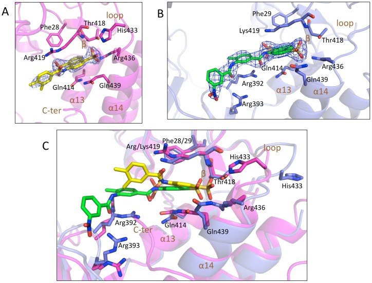
Figure 3. Interaction network of hNV and mNV-RdRp/6 complex.
A) Fragment of 6 (carbon atoms in yellow) bound to hNV-RdRp in cartoon (magenta). All the amino acids involved in interaction with the inhibitor molecule are shown in sticks (carbon atoms in magenta). B) 6 (carbon atoms in green) bound to mNV-RdRp in cartoon (blue). All the amino acids (carbon atoms in blue) involved in interaction with the molecule are shown in sticks. 2Fo-Fc electron density contoured at 1 sigma in blue grid. C) Superposition of hNV-RdRp and mNV-RdRp, showing the structures of 6 (carbon atoms in yellow/green, respectively) bound to hNV-RdRp and to mNV-RdRp (cartoon in magenta/blue, respectively). The interacting amino acids are shown in sticks (magenta/blue carbon atoms, respectively).

