
Figure 1. CD133-positive cells line hemangioma vessels.
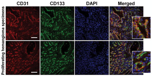
Proliferating hemangioma specimens were double-labeled for CD31 (endothelial cell marker; Red) and CD133 (stem cell antigen; Green). DAPI (blue) was used as counterstain. Staining illustrates complete co-localization of CD133 and CD31 in both proliferating hemangioma specimens (images were taken at 20X magnification; inserts illustrate high magnification; scale bar = 200μm).