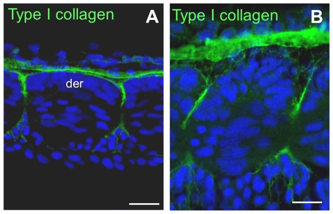
Figure 1. Collagen I is concentrated at the surface of the somites.
(A and B) Frontal sections through the trunk of a 13 dpf trout embryo. (A) Posterior tail. Collagen I immunofluorescence localises to the anterior and posterior edges and at the lateral surface of the dermomyotome. (B) Anterior tail. Collagen I immunofluorescence is present along the space separating adjacent somites. der: dermomyotome. Scale bars in A and B, 15 μm.

