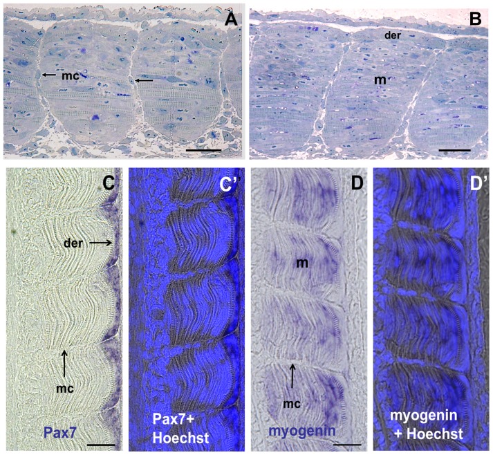
Figure 2. The intermyotomal space is colonized by fibroblast-like cells that do not express myogenic markers.
(A and B) Semithin frontal sections through the trunk of an eyed stage (17 dpf) embryo with final number of somites and pigmented eyes. (A) Posterior tail. Myoseptal cells are visible in the medial half (arrows) of the space separating two somites. (B) Anterior tail. Myoseptal cells are visible throughout the medio-lateral extent of the intermyotomal space. (C) Pax 7 expression is restricted to cells forming the dermomyotome-like epithelium at the surface of the myotome. (C′) Merged image showing Pax7 labeling and Hoechst staining for nuclei visualisation. (D) Myogenin expression is observed in the primary myotome located below the dermomyotome-like (D′) Merged image showing myogenin labeling and Hoechst staining. mc: myoseptal cells, m: myotome, der: dermomyotome. Scale bars in A, B, C and D, 25 μm.

