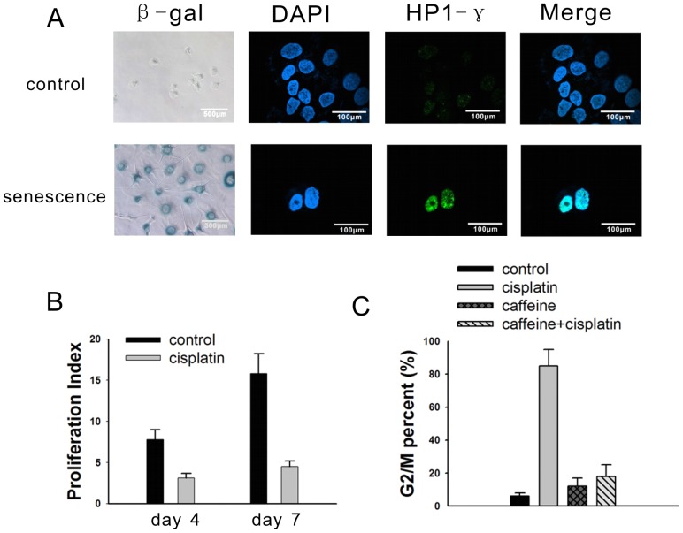
Figure 1. Cisplatin induces a senescent phenotype in NG108-15 cells.
(A) Untreated cells and cells treated with 5 µg/ml cisplatin were stained with β-gal at pH 6.0 (200×, light microscopy), DAPI (600×, confocal microscopy), and immunofluorescent HP1-γ protein (600×, confocal microscopy). The DAPI and HP1-γ stains were merged. (B) The cells were labeled by CFSE to determine the proliferation index on day 4 and day 7 following cisplatin treatment. The proliferation index of the untreated cells on 0 hour was 1. (C) 7 days after treatment with 5 µg/ml cisplatin, 5 mmol/L caffeine, or a combination of cisplatin and caffeine, the NG108-15 cells were harvested for FACS analysis. The results are the mean of 3 independent experiments.

