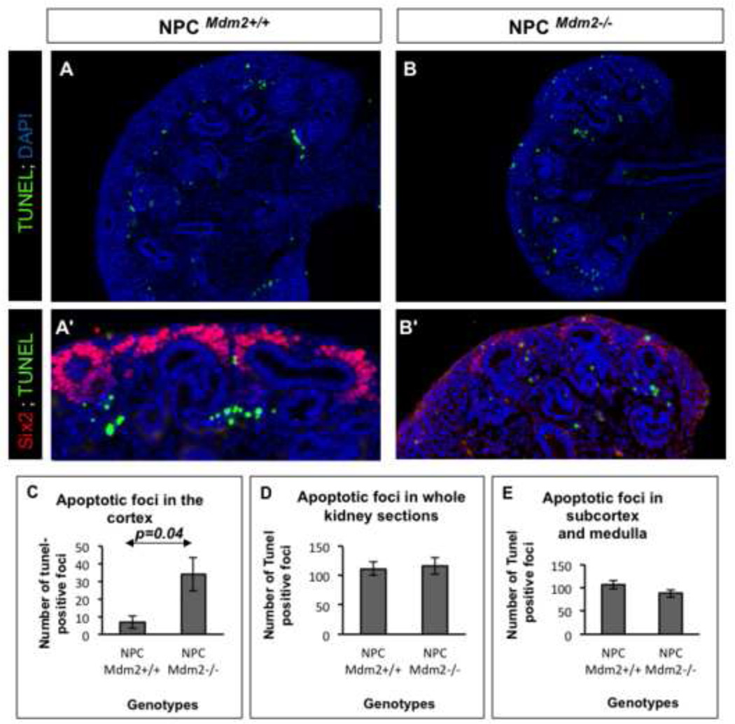
Figure 5. (A–E). The NPCMdm2−/− kidneys show significant elevation in apoptosis by TUNEL assay.
Sections of E14.5 NPCMdm2+/+ kidneys (A) show largely subcortical apoptotic foci. By contrast, in the NPCMdm2−/− kidneys (B) there are numerous apoptotic foci in the cortical, nephrogenic zone including the dorsal aspect of ureteric bud tips. (A', B') Panels showing wild type and mutant kidney sections co-stained for Six2 following TUNEL assay to highlight the presence of apoptotic cells in the cap mesenchyme of only the latter. (C) A graph showing a significant increase (4.9 fold, p=0.04) in TUNEL-positive apoptotic foci in the cortical regions of NPCMdm2−/− kidneys relative to control kidneys. (D) Counts of apoptotic foci in complete kidney sections (cortex, sub-cortex, and medulla) or in regions interior to the cortex remain unchanged or show an insignificant drop in apoptosis. (n= 7 animals per genotype)

