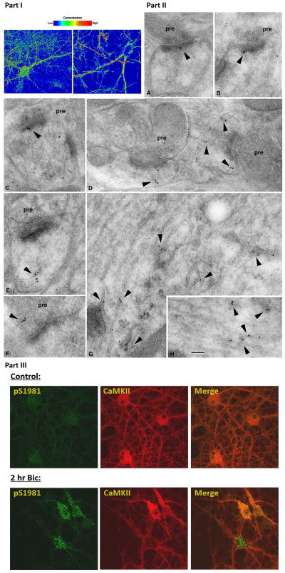
Figure 5. ATM localization and activation in neurons.
Part I: Immunofluorescence localization of ATM in mixed hippocampal/cortical neurons: two different antibodies were utilized to investigate the localization of ATM in neurons (note: z-section analysis of neurons revealed substantial nuclear localization as well, see supplementary movie file). Part II: Immuno-gold electron micrograph study of ATM in the hippocampus; Labeling (arrowheads) is associated with vesicles or tubulovesicular structures in postsynaptic spines (A–E) and also in presynaptic terminals (F). In dendrites, labeling often is concentrated on tubulovesicular structures (G, H) including those associated with distinct endosomal complexes (G; straighter, regular tubular structures in G and H are microtubules running parallel to the dendrite axis). Micrographs were taken in the CA1 stratum radiatum (A, B, E, G) and pyramidale (D, H) and the CA3 stratum lucidum (C, F). pre=presynaptic terminal. Scale bar is 100 nm. Part III: ATM activation by synaptic activity. ATM pS1981, an autophosphorylation site on ATM, is determined in primary neurons treated without (control) or with (Bic) 2 hours of bicuculline. CamKII costaining indicates excitatory neurons.

