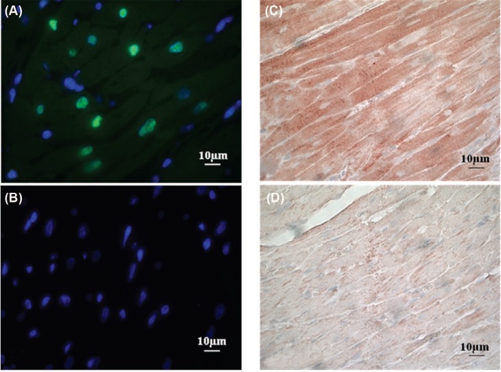
Figure 6:
Immunohistochemical verification of cardiac radiation exposure after a total dose of 21 Gy. (A) Representative image of the immunofluorescence staining of γH2Ax (green) with DAPI counterstain of nuclei (blue) in the heart at 2 hours after irradiation. (B) Representative image of the immunofluorescence staining of γH2Ax with DAPI counterstain of nuclei in the heart at 2 hours after control treatment (sham-irradiation). No γH2Ax was detected after sham-irradiation. (C) Representative image of the immunohistochemical staining of nitrotyrosine in the heart at 6 hours after irradiation. (D) Representative image of the immunohistochemical staining of nitrotyrosine in the heart at 6 hours after sham-irradiation. Minimal nitrotyrosine was detected after sham-irradiation.

