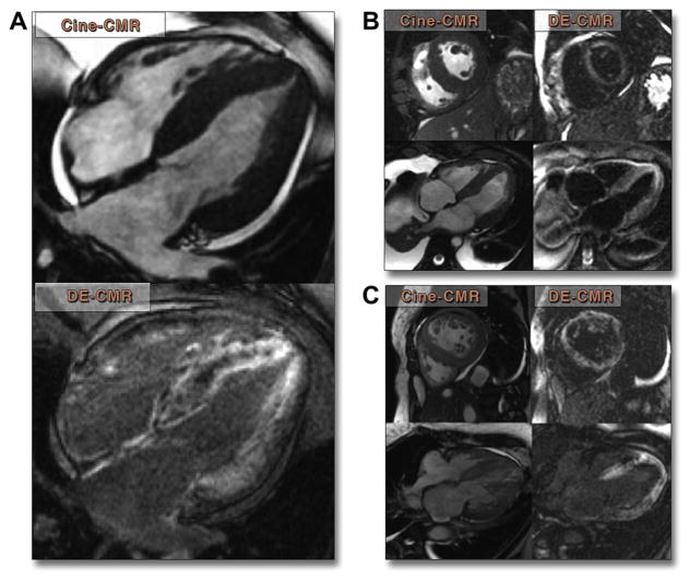
Figure 3. Typical Findings in 3 Patient Examples.
(A) Four-chamber cine cardiac magnetic resonance (CMR) and delayed-enhancement cardiac magnetic resonance (DE-CMR) images in a 52-year-old man with systemic primary amyloidosis 5 months prior to his death. Diffuse, primarily subendocardial hyperenhancement is seen in both the left and right ventricles as well as in the interatrial septum. (B) A 61-year-old man with myeloma and new-onset heart failure showing diffuse subendocardial hyperenhancement. Patient died 6 months later. (C) A 49-year-old woman with restrictive cardiomyopathy by echocardiography and invasive hemodynamics showing diffuse hyperenhancement of the left ventricle. Patient died 45 days later.

