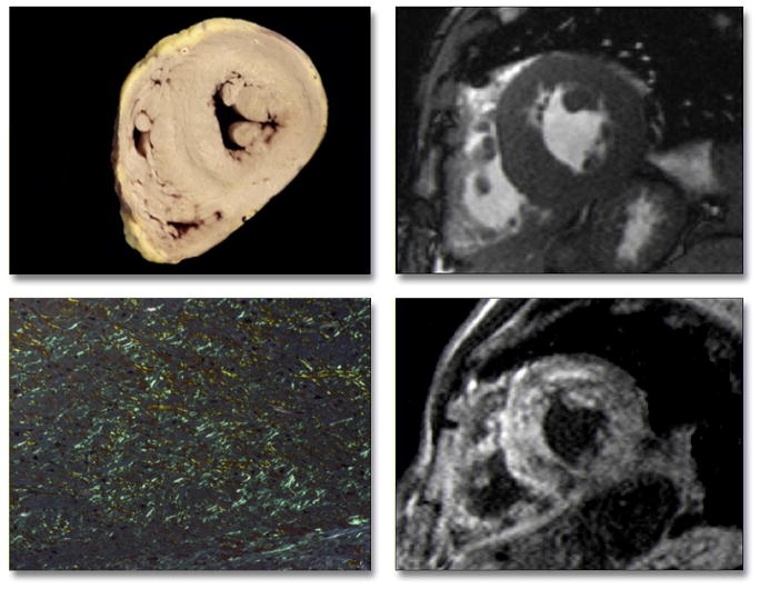
Figure 5. Pathology Correlation in a Patient With Primary Amyloidosis.
Gross morphology (top left) shows globally-increased wall thickness of both the left and right ventricles, as also demonstrated in vivo by cine cardiac magnetic resonance (top right). Tissue from the interventricular septum stained with Congo red shows marked interstitial amyloid deposition, with apple-green birefringence under polarized light (bottom left) correlating with diffuse hyperenhancement on delayed-enhancement cardiac magnetic resonance, which was particularly evident in the septum (bottom right).

