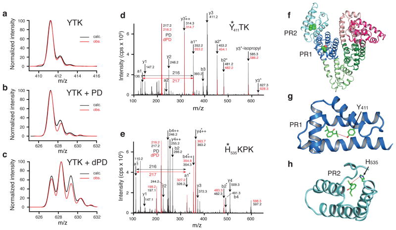
Figure 2. Labeling human serum albumin with ortho-propofol diazirine.
Calculated and observed isotopic distributions for (a) unlabeled YTK peptide, (b) YTK peptide labeled with protiated ortho-propofol diazirine and (c) YTK peptide labeled with partially deuterated ortho-propofol diazirine. MS2 data for peptides YTK (d) and HKPK (e) with protiated ortho-propofol diazirine (black) and deuterated ortho-propofol diazirine (red). The asterisks denote the presence of photolabel bound to the peptide fragment. The two sites are illustrated in (f) with PR1 being in subdomain IIIA (dark blue) and PR2 being in subdomain IIIB (light blue). More detailed views are shown in (g) for PR1 where the labeled Y411 is shown and in (h) for PR2 where the labeled H535 is shown. Dashed lines mark the distances between the propofol molecules to Y411 (4.9 A) and to H535 (5.0 A).

