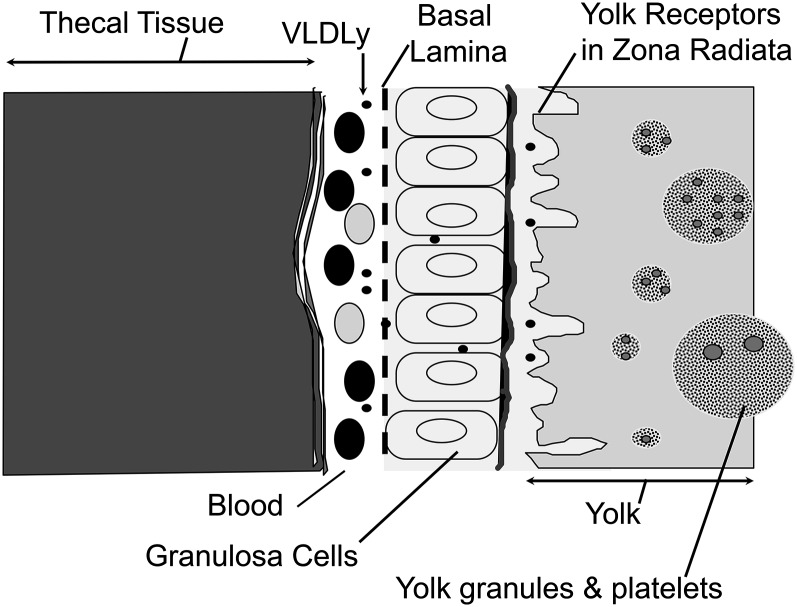
FIGURE 1.
Diagram of avian yolk follicle in cross-section. Note that ovarian tissue layers (theca and granulosa) are vascularized, providing the means for delivery of hepatic lipoproteins to the oocyte surface (zona radiata) in which lipoprotein receptors are located. Yolk forms by receptor-mediated uptake of intact VLDL and vitellogenin. Only yolk-targeted VLDL, 25–30 nm in diameter and resistant to hydrolysis by lipoprotein lipase, passes through the granulosa basal lamina; vitellogenin is a large phospholipidated protein ∼10 nm in diameter. VLDLy, yolk-targeted VLDL.

