
Fig. 5.
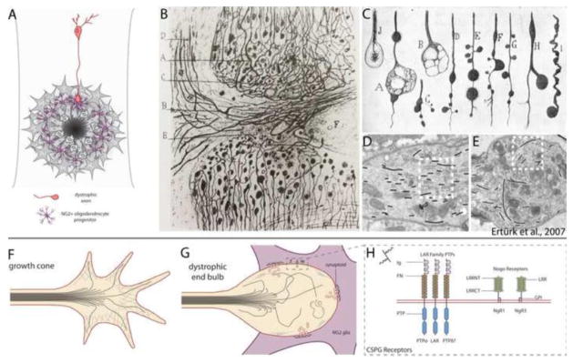
The glial scar acts as the primary barrier to regenerating axons. (A) Regenerating axons halt abruptly at the glial scar in close association with NG2 glia, and axon endings take on a state of dystrophy. (B, C) Reprinted from Cajal’s Degeneration and Regeneration of the Nervous System (1928) by permission of Oxford University Press, USA. (B) Drawing by Ramón y Cajal of the edges of a complete transection lesion to the spinal cord. Cajal noted that adjacent to the lesion, fine axons terminate in rings or little clubs while larger axons end in voluminous clubs. (C) Several drawings by Cajal of the various appearances of retraction clubs at the lesion. (D, E) Data republished from Ertürk et al. (2007) with permission from the Society for Neuroscience; permission conveyed through the Copyright Clearance Center, Inc. (D) Electron micrograph of a growth cone in the lesioned sciatic nerve. Growth cones exhibit highly parallel arrays of microtubules (traced with black lines). (E) Electron micrograph of a retraction bulb from a lesioned central axon of a dorsal root ganglion neuron. Retraction bulbs exhibit disorganized or splayed microtubules. (F) Illustration of a growth cone. Microtubules are bundled and oriented toward the direction of the growing axon. Growth cones also have a highly organized F-actin network and several filopodia. (G) Dystrophic end bulbs have disorganized microtubules and a disrupted F-actin network. Large membrane blebs or inclusions can be observed within retraction clubs. Dystrophic axons associate with NG2 glia in the outer margin of the lesion core, forming “synaptoids” that exhibit similar properties to a mature synapse, with numerous presynaptic vesicles and omega structures, and a post-synaptic density at the NG2 glial membrane. (H) Several recently identified CSPG receptors act to signal inhibition, and may entrap axons on the surface of NG2 glia. These receptors fall into two classes: the LAR family of transmembrane protein tyrosine phosphatases (which includes PTPσ, LAR, and PTPδ), and the Nogo receptors NgR1 and NgR3. LAR family receptor protein tyrosine phosphatases have high sequence similarity and contain three N-terminal Ig domains, 8 fibronectin repeats, and two intracellular tandem phosphatase domains. The first Ig domain of PTPσ, LAR, and PTPδ contains a canonical GAG binding motif (BB-X-BB, where B is lysine or arginine). NgR1 and NgR3 have several leucine-rich repeats, are GPI-anchored, and contain a cluster of basic residues in the C-terminal stalk required for binding CSPGs.