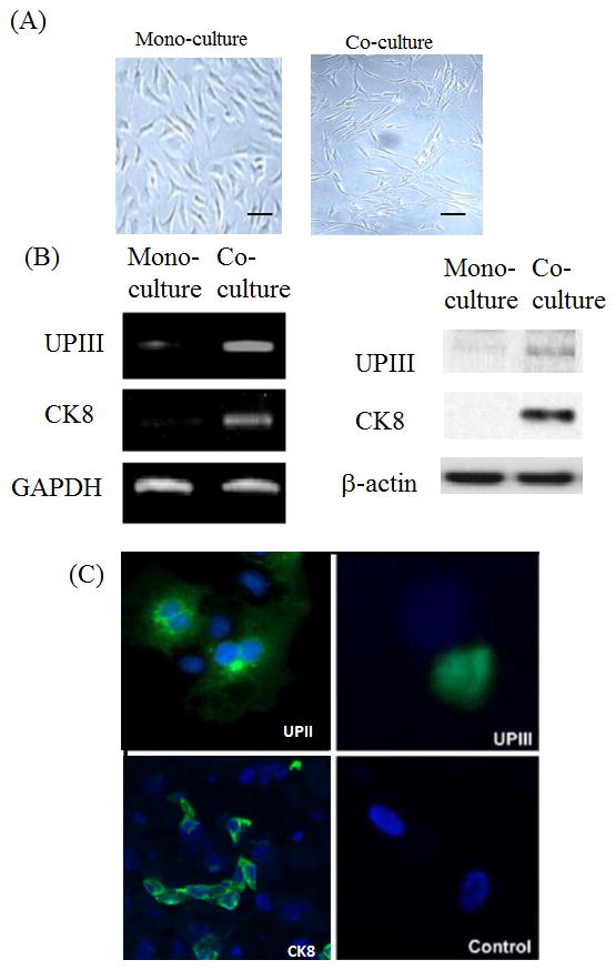
Figure 1.

(A) Human amniotic fluid stem cell morphology from the mono-culture and co-culture. Human stem cells were culture in the mono-culture and co-culture systems and morphology was observed after 2 weeks. Scale bar 100 μm. (B) Left panel: RT-PCR was performed with FGF10, uroplakin III and cytokeratin 8 gene primers in stem cells co-cultured with bladder cancer cell line LD605. Right panel: Western blot was performed with the stem cells co-cultured and mono-cultured. FGF10 protein and urothelial markers, UPIII and CK8, were examined on the PAGE analysis. (C) Immunofluorescence of bladder markers uroplakin II, uroplakin III and cytokeratin 8 in the co-culture system. Amniotic fluid stem cells cultured alone (mono-culture) served as a control.
