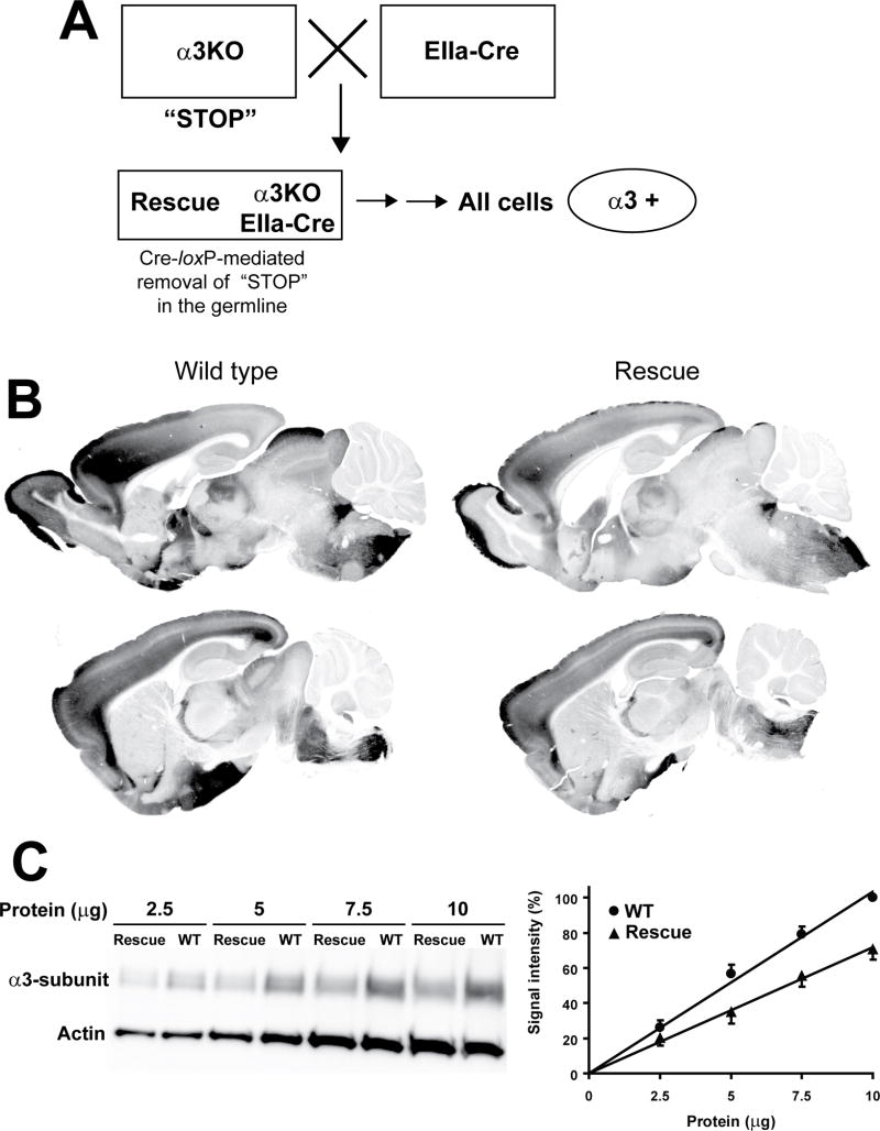
Figure 4. Localization and quantification of the α3 knockout in global rescue mice.
A. Generation of global Rescue mice. An α3KO mouse (with artificial exon 5, the “STOP” signal) is bred with an EIIa-Cre mouse19. Some of the offspring will carry both the α3KO allele and the EIIa-Cre transgene. This mouse is bred with a wild type mouse to breed out the EIIa-Cre transgene (depicted by two sequential horizontal arrows) to confirm that the artificial exon 5 (the “STOP” signal) has been removed from the germline, so that the α3 subunit is expected to be expressed in all cells in which it is naturally expressed.
B. Immunoperoxidase staining of perfusion-fixed parasagittal brain sections: α3 subunit distribution pattern is equivalent in wild type and global rescue mice. Representative sections from 2 wild type and 2 global rescue mice are shown.
C. Left panel: Representative Western Blot: α3 subunit expression level is decreased in global rescue mice compared to wild type mice. Right panel: Quantification of Western Blot signal: signals were normalized to the α3 subunit signal at 10 μg protein in wild type mice (100%). α3 subunit expression level in global rescue mice is 70±7% of wild type expression. Data represent the mean±SEM of five experiments.

