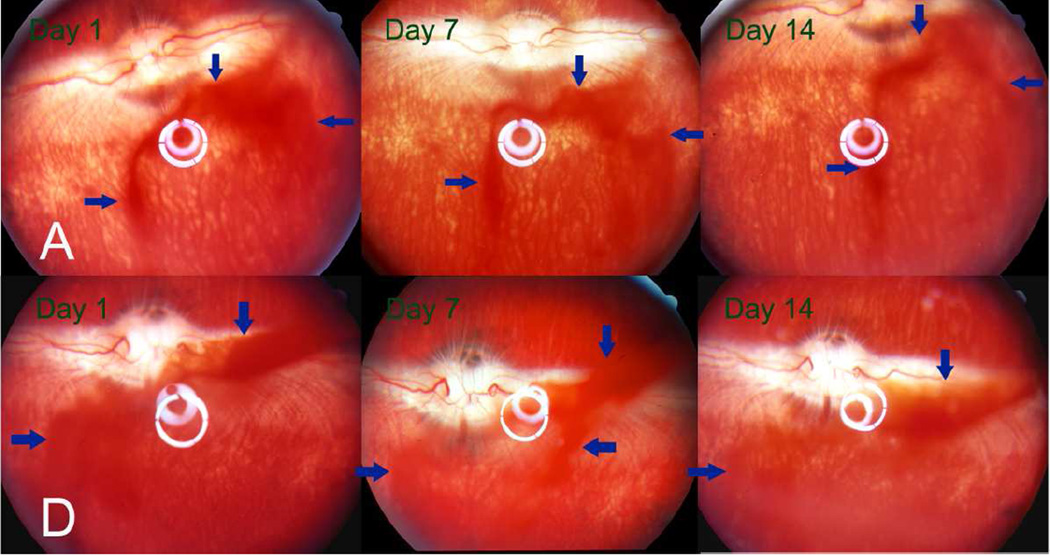
Figure 7.
Fund us images of the rabbits’ eyes at observation time points 1, 7 and 14 days after injection of 3 mg particle A (upper panel) or particle D (lower panel) into the right eye of the rabbits. The DNR-loaded particles appeared reddish (arrows) due to the red color of the drug. The quantity of particles in the vitreous appeared to have decreased in both groups at 2 weeks after injection. The vitreous, retina, and optic nerve head appeared normal.

