
Figure 2.
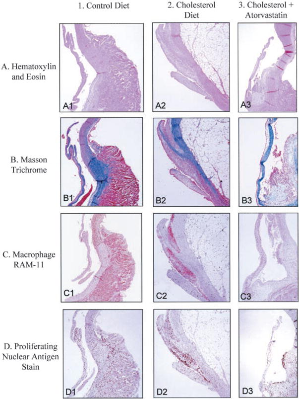
Light microscopy of rabbit aortic valves and aorta. Left column, control diet; middle column, cholesterol diet; right column, cholesterol diet plus atorvastatin. In each panel, aortic valve leaflet is positioned on the left, with aorta on the right. All frames ×12.5 magnification. A, Hematoxylin and eosin stain; B, Masson trichrome stain; C, macrophage, RAM 11; D, PCNA stain.