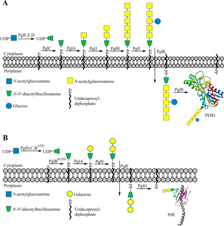
Figure 1.
(A) N-linked protein glycosylation pathway from C. jejuni showing the heptasaccharide glycan attached to the PEB3 protein. (B) O-linked protein glycosylation pathway from N. gonorrhoeae showing the trisaccharide glycan attached to the PilE protein. Both pathways utilize the unique, bacterial sugar diNAcBac at the reducing end of the glycan. Abbreviations: ATD, acetyltransferase domain; PGTD, phosphoglycosyltransferase domain.

