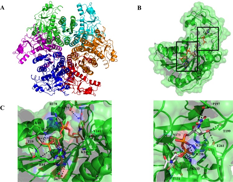
Figure 8.
(A) Ribbon representation of the H. pylori dehydratase PseB biological hexamer unit with each protomer individually colored for the sake of clarity. (B) Ribbon and space-filling representation of the monomer bound to UDP-GlcNAc (gray) and NADP+ (brown) (PDB entry 2GN6). (C) Major PseB active site interactions depicted as dashed lines with NADP+ (left) and UDP-GFlcNAc (right).

